
04 March 2026 | Øyvind Rørbakken, TU Delft, Delft, The Netherlands | Blog
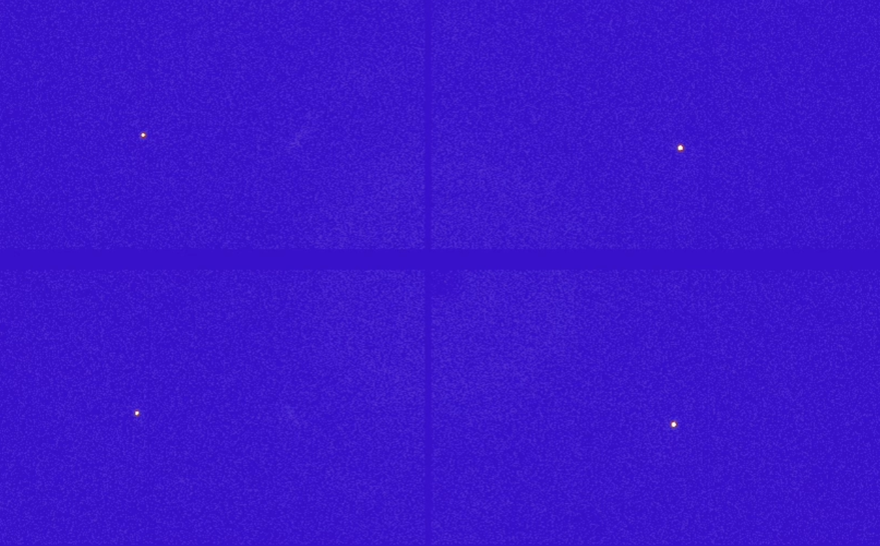
Look at the animation below. What do you see? It may seem like tiny stars twinkling in the night sky. They appear and disappear, in a somewhat symmetrical pattern. Why is that?
Imagine a dancer with a sequined outfit, on a stage in the spotlight. The dancer does a pirouette. Watching from the audience, you might at some points see extra bright glimpses of light scattered from the spotlight to your eye by the sequins on their outfit.
What we observe here is not so different. This animation was made from a series of detector images from an x-ray diffraction (XRD) experiment. The sample, a small, single crystal rod, rotates like a dancer doing a pirouette on stage in the spotlight from an intense beam of x-rays from a beamline at the ESRF synchrotron. The detector spans a plane surface, and the bright spots we see are located where the scattered x-rays hit the detector.
This happens due to the phenomenon of diffraction. Most of the intense, high energy x-ray beam passes straight through the sample, but parts of the beam can scatter from the atoms within. This sample was an ordered structure, a crystal, of the elements manganese, iron, phosphorous and silicon. For some positions on the detector, the distance to a collection of atoms in the crystal happens to match particularly well with the wavelength of the x-rays. This causes a local pile-up of x-ray intensity at the detector and appears as bright spots standing out from the rest of the detector area.
The diffraction spots may carry surprisingly rich information about the internal structure of the sample. The condition for diffraction changes as the sample is oriented at different angles, but for some orientations of the sample, you may observe the same diffraction pattern. This points to the fact that the structure has some internal symmetry, where a certain rotation leaves the crystal looking unchanged. With high resolution diffraction data, you can characterize internal symmetries of the structure, distances between atoms and even the type of element sitting at specific positions in the structure. Diffraction techniques are among the most widely used characterization techniques used in science concerned with understanding the atomic and molecular world.
Putting samples in various situations, say under heating or increasing the strength of an applied magnetic field, while observing diffraction patterns can also unveil the microscopic responses in materials in operation. The information from such in-situ experiments is useful for both fundamental science and for guiding the future iterations and developments of materials and devices by acquiring information that would otherwise be unattainable to us by just studying a sample before and after operation.
Microscopic characterization experiments like these are what I am concerned with in the HEAT4ENERGY project. Knowledge about the correlations between the magnetic properties of the thermomagnetic materials at a large scale and other structural properties at a microscopic scale is important for understanding the strengths and weaknesses of the materials, and it guides the selection of materials well suited for specific applications and devices going forward.
Attribution: Originally published by HEAT4ENERGY. Reposted with permission. Original article: https://heat4energy.eu/blog/blog-7-what-can-microscopic-characterization-teach-us-about-thermomagnetic-materials


